
Hello everyone!
When people think of ultrasounds, many think of the black and white grainy photo of a baby on a little slip of paper. However, as you see from previous post, ultrasounds have an integral role in emergency medicine too.
Because I've gone over the emergency ultrasounds in previous posts, I want to talk more about the role of ultrasounds in prenatal care. Plus, who doesn't want to see cute little babies?
For some background, a woman will normally have around two ultrasounds, one during her first trimester and another one at around 18 weeks-22 weeks pregnant (about midway through a woman's pregnancy). A gestational sac can be identified through an ultrasound as early as 4 1/2 weeks and even more mind blowing, a fetal heartbeat can sometimes be detected as early as 5- 6 weeks!
The ultrasound done during the first trimester is called a 'basic level one ultrasound' and is used to:
- confirm a fetal heart
- make sure the fetus is attached to the uterus
- date a pregnancy
The second ultrasound that is done midway during a pregnancy is called a 'level 2 ultrasound' and can give a detailed image of the growing fetus. The actual image that is projected from the ultrasound and the image a mother takes home to show her friends and family is called a Sonogram. The medical reasoning behind these freeze-framed images is because babies this late into trimester move a lot so the physician takes these snapshots to more accurately check that the baby is developing normally.
Sometimes, if an physician is unable to clearly see if the baby is developing normally, or the mother's had previous complications in pregnancy, she will often have multiple ultrasounds as part of her prenatal care plan.
That's about everything I wanted to cover so here are some pictures to wrap this post up:
First Trimester:
 |
| Week 4: Gestational Sac |
 |
| Week 5: Gestational sac and yolk sac of twins! the gestational sac is the back and the little circle within the black is a yolk sac (provides nutrients to fetus). |
 |
| Week 9 |
 |
| Week 12 |
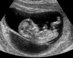
Second Trimester:
 |
| Week 16 |
Third Trimester:
 |
| Week 29. The baby's bones and muscles are growing stronger. |
 |
| Week 30: Baby's eyes are opening and further developing facial features. |


0 comments:
Post a Comment